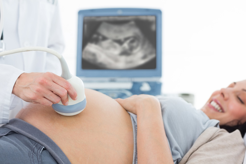
Ultrasound

Obstetrics
1st Trimester Scans – Dating Scan
A dating scan is performed around week 8 of pregnancy but can be done as early as 5 weeks. It is used to confirm due dates, assess the viability of the pregnancy, check the number of embryos, provide maternal reassurance, and to rule out ectopic pregnancy (fetus developing outside of the womb).
1st Trimester Scan – Nuchal translucency
A nuchal translucency (NT) scan is a screening test that assesses whether or not your baby is likely to have Down syndrome.
Nuchal translucency is a collection of fluid under the skin at the back of the baby’s neck. It can be measured using ultrasound: between 11 weeks and 13 weeks of pregnancy. All babies have some fluid at the back of their neck. But many babies with Down syndrome have an increased amount.
Before this the scan you need to have a blood test to check your PAPPA and bHCG levels is needed to calculate the risk factor. You need to bring these results to your appointment or ask your pathology centre to send them through to Northamultrasound.
12-16 Week Early Anatomy Scan
Advances in ultrasound technology mean we can start to examine your baby’s anatomy much earlier than in the past. An early fetal anatomy scan at around 13-16 weeks can help to identify problems, this does not take the place of the 20 week anatomy scan and is best for
- Women who have not had a scan earlier in the pregnancy
- Couples with a previous child affected by a structural anatomical problem
- Twin and triplet pregnancies
- Women with chronic medical problems, such as diabetes, or using certain medications
2nd trimester scans – Anatomy Scan
Around week 20 of pregnancy, an anatomy scan is performed to rule out abnormalities that can be visualised with ultrasound imaging such as cleft lip, spina bifida, heart defects, and many other abnormalities. It is also used to check that the fetus size is within normal limits and record the location of the placenta. The gender of the fetus can also usually be established during this scan.
3rd Trimester Scans – Growth Scan
Usually conducted from week 28 of pregnancy, the growth scan is used to check the baby’s growth by measuring its head, abdomen, and thigh bone. It is also used to assess the amount of amniotic fluid surrounding the baby and record the position of the placenta.
Gynecology
Pelvic Ultrasound (female)
An ultrasound scan is used to view the uterus (womb) and ovaries for things such as ovarian cysts, endometrial disorders, fibroids, Intrauterine Contraceptive Device (IUCD) location, or other disorders for pelvic pain and other symptoms.
Follicle Tracking
Ultrasound can be used to help your doctor manage fertility problems by tracking the size and stage of follicles on the ovaries. The uterus and the inside lining (endometrium) can be checked and measured to assessed with fertility.
Pelvic Prolapse
Ultrasound can be used to assess the movement of the pelvic organs to assess for prolapse.
General Ultrasound
Abdominal Ultrasound
An ultrasound scan is used to assess the pancreas, aorta, liver, gallbladder, bile ducts, kidneys and spleen. It is also used to check for fluid in the abdomen (ascites). Gallstones, liver and kidney cysts, aortic aneurysms and other abnormalities can be assessed with ultrasound.
Renal Ultrasound
An ultrasound scan is used to assess the urinary bladder and kidneys to check for stones, blockages, lesions and other abnormalities.
Thyroid Scan
An ultrasound scan of the thyroid is used to check for cysts, nodules, and abnormalities and to assess the size and offer follow-up assessment.
Breast Scan
An ultrasound scan of the breast is used to assess a palpable lump or to monitor the size of previously seen lesions.
Scrotal Scan
An ultrasound scan of the scrotum is used to assess a palpable lump, testicular pain, epididymal lumps or abnormalities. It is also used to assess the scrotal size and to rule out testicular torsion, hydrocele and varicocele.
Lumps and bumps
An ultrasound scan is also used on a variety of palpable lumps and bumps to assess their nature.
Foreign Body Ultrasound
An ultrasound scan is used to check for the presence of a foreign body and mark the location for removal if required.
Musculoskeletal
Shoulder Scan
An ultrasound scan of the shoulder is used to check for muscle tears and fluid associated with the rotator cuff, swelling and subluxation of the acromioclavicular (AC) joint, and the long head of the biceps tendon (LHB) can be assessed. Frozen shoulder and other joint pathology can also be assessed with ultrasound.
Ankle Scan
An ultrasound scan of the ankle is used to assess tendons and ligaments for damage and also the ankle joint for fluid. Ruptures, tears, and tendinopathy of the Achilles tendon can also be well assessed with ultrasound imaging.
Knee Scan
An ultrasound scan of the knee is used to assess the Medial Collateral Ligament (MCL), Lateral Collateral Ligament (LCL), quadriceps and patella tendon for strains and tears. The popliteal fossa is also checked for Baker’s cysts, Deep Vein Thrombosis (DVT) and other abnormalities
Elbow Scan
An ultrasound scan of the elbow is used to rule out lateral epicondylitis (tennis elbow) and medial epicondylitis (golfer’s elbow) and assess injury to the lower parts of the biceps and triceps tendon. The joint fluid and surrounding soft tissues is also assessed.
Wrist Scan
An ultrasound scan of the wrist is used to check for swelling of the tendons in the wrist and joint fluid. De’Querveins abnormality is a common condition of the wrist that is diagnosed with ultrasound imaging. The nerves within the wrist can also be assessed for carpal tunnel pathology.
Muscle Scans
Muscle tears are common and can be well visualised with ultrasound, muscle tears and strains to the hamstrings, quadriceps and calf muscles are some common regions of injury.
Appointment Enquiries
Staff will contact you to confirm
"*" indicates required fields